UPMC has conducted several neurosurgical case studies using the Neuroendoport procedure. Learn how UPMC neurosurgeons have removed deep brain tumors and cysts through the Neuroendoport — a narrow tube the circumference of a dime. This innovative technique minimizes trauma to the brain and surrounding nerve tissues.
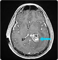
Neuroendoport Surgery for Colloid Cyst
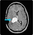
With a large colloid cyst of the third ventricle, a 41-year-old veteran suffered from unremitting headaches, confusion, and fainting spells. Read how UPMC surgeons used Neuroendoport surgery to completely remove the cyst and bring immediate relief to the patient.
» Read full case study
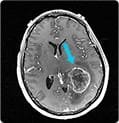
Neuroendoport Surgery for Glioblastoma
With a lesion covering a large amount of functional brain cortex, critical for speech function, a woman faced standard brain surgery requiring a large opening in the dura (brain covering), and lowering her chances of ever regaining speech function. Read how UPMC surgeons used Neuroendoport surgery to remove the 4 cm glioblastoma, restoring speech function.
» Read full case study
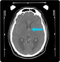
Neuroendoport Surgery for Metastatic Breast Cancer to the Frontal Lobe
With a tumor in her brain’s deep frontal region, a 52-year-old woman suffered from weakness and speech problems. A large opening or brain dissection would risk worsening of her weakness and an inability to recover her speech. Read how UPMC surgeons used Neuroendoport surgery to remove the tumor and how the patient recovered.
» Read full case study
With an intraventricular tumor nearly two inches in diameter, a 47-year-old man suffered from relentless headaches, confusion, and visual disturbances. Read how UPMC surgeons used Neuroendoport surgery, with minimal manipulation of the surrounding brain, to relieve the patient’s symptoms and preserve his speech and vision.
» Read full case study
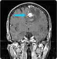
Neuroendoport Surgery for Recurrent Intraventricular Meningioma
With a tumor the size of a racquetball in her ventricle, a 45-year-old woman suffered progressive headaches and numbness on the left side of her body. Read how UPMC surgeons combined Neuroendoport surgery with radiosurgery to return the patient to normal function.
» Read full case study