Neuroendoport Clinical Case Study
The Patient
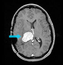
A 45-year-old woman who had surgery six years prior for an intraventricular meningioma developed progressive headaches and numbness on the left side of her body. MRI scans showed a massive recurrent meningioma, the size of a racquetball.
|
Pre-surgical scan shows a massive 6cm meningioma in the ventricle.
|
|
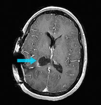
Post-surgical scan shows the successful removal of the intraventricular meningioma.
|
The Challenge and Solution
The 6 cm tumor was highly calcified, like a rock. Using the Neuroendoport technique, the tumor was removed in two stages on separate days, except for a few millimeters of residual tumor, which was subsequently treated with radiosurgery. The patient resumed normal function following resection, and no increased trauma to the brain was incurred by the use of the Neuroendoport technique.
The Result
The post-surgical MRI shows complete removal of the meningioma. The patient's symptoms completely resolved. Treatment and results may not be representative of all similar cases.
Treatment and results may not be representative of all similar cases.