Clinical Case Study
The Patient
A 17-year-old girl came to the hospital with headaches after a fall. The CT scan and MRI showed a large trigeminal schwannoma at the base of the skull.
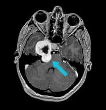
Pre-surgical scan shows large tumor at the base of the skull.
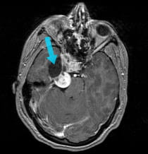
Scan after first surgical procedure shows front half of tumor has been removed using EEA surgery.
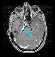
Scan after second surgical procedure shows complete tumor removal.
The Challenge
The tumor had two portions, separated by the petrous bone and the brain covering. The petrous bone contains many important structures, including critical cranial nerves and the carotid artery.
The Solution
To avoid any damage to the nerves in and around the petrous bone and the carotid artery, the UPMC surgeons performed two separate surgeries from different angles to remove the trigeminal schwannoma.
The Result
Surgeons were able to completely remove the trigemina schwannoma. The girl temporarily had double vision that completely resolved over time, and some mild numbness related to the nerve from which the tumor originated.
Treatment and results may not be representative of all similar cases.


