Learn how UPMC neurosurgeons have used the Endoscopic Endonasal Approach (EEA) to remove difficult tumors and lesions from the skull base — through the nose and sinuses.
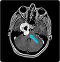
Trigeminal Schwannoma
Scans of a 17-year-old girl who complained of headaches showed a large trigeminal schwannoma at the base of the skull. The tumor had two portions, separated by the petrous bone and the brain covering. Read how UPMC surgeons were able to avoid any damage to the nerves in and around the petrous bone and the carotid artery by using several techniques, including EEA, to completely remove the tumor.
» Read full case study
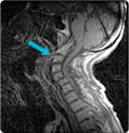
Arthritic Breakdown in Upper Cervical Spine
An elderly woman with arthritic breakdown at the top of her spine may have faced traditional surgery, including a tracheotomy and an incision in the back of the throat that would have kept her from eating right after surgery and potential long-term problems with swallowing. Read how UPMC surgeons performed surgery using EEA to remove the bone that was pressing on the patient's spinal cord, which allowed the patient to start eating the day after surgery.
» Read full case study
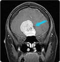
Olfactory Groove Meningioma
Traditional surgery for an olfactory groove meningioma would require a long scalp incision and lifting the front of the brain to get to the tumor. In the case of a 35-year-old woman experiencing headache and vision loss from pressure and swelling in her brain, the tumor, because of its height, required a long reach to the top of the tumor. Read how UPMC surgeons used EEA to remove the tumor without lifting the brain.
» Read full case study


