5/18/2020
 PITTSBURGH – Neuroscientists at the University of Pittsburgh Brain Institute have traced neural pathways that connect the brain to the stomach, providing a biological mechanism to explain how stress can foster ulcer development.
 “Pavlov demonstrated many years ago that the central nervous system uses environmental signals and past experience to generate anticipatory responses that promote efficient digestion,” said Peter Strick, Ph.D., Brain Institute scientific director and chair of neurobiology at Pitt. “And we have long known that every increase in unemployment and its associated stress is accompanied by an increase in death rates from stomach ulcers.”
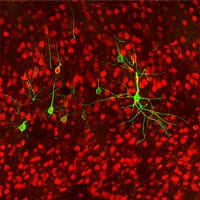
 Strick and Levinthal found that the parasympathetic — “rest and digest” — nervous system pathways trace back from the stomach mostly to a brain region known as the rostral insula, which is responsible for visceral sensation and emotion regulation.
 These insights also could change clinical gastroenterology practice. Knowing that the brain exerts physical control over the gut gives doctors a new way to approach bowel problems.
IMAGE INFO: (click images for high-res versions)
1st:
CAPTION: Neurons Projecting to the Stomach: Rabies-virus-labeled neurons (green) in the insula that send projections down to the stomach.
CREDIT: David Levinthal, M.D., Ph.D., and Peter Strick, Ph.D.
2nd:
CAPTION: Peter Strick, Ph.D., Brain Institute scientific director and chair of neurobiology, University of Pittsburgh
CREDIT: University of Pittsburgh
3rd:
CAPTION: Two Nervous Systems: Two opposing circuits connecting the brain to the stomach convey either "fight or flight" or "rest and digest" commands to the gut.
CREDIT: David Levinthal, M.D., Ph.D., and Peter Strick, Ph.D.
4th:
CAPTION: David Levinthal, M.D., Ph.D., assistant professor of gastroenterology, hepatology and nutrition, University of Pittsburgh
CREDIT: UPMC


