8/5/2022
In a study, published today in Science Immunology, the team profiled molecular features of T cells as they progressed from early to terminal exhaustion in a mouse model of melanoma. They unexpectedly found that even the most terminally exhausted T cells retain some capacity to be functional again and identified approaches to overcome exhaustion, opening potential new avenues for cancer immunotherapy.
Over time, tumor-fighting T cells may enter the early phase of exhaustion, a progenitor-like cell, which eventually differentiates into poorly functional, terminally exhausted cells. While current cancer immunotherapies can be successful at reversing exhaustion in these progenitor cells, giving T cells renewed energy to attack cancer cells, those that are terminally exhausted tend not to respond to such therapy.
 “To bring the promise of immunotherapy to more cancer patients, we need to uncover more about why T cells fail,” said co-senior author Greg Delgoffe, Ph.D., associate professor of immunology at Pitt and director of the Tumor Microenvironment Center at UPMC Hillman Cancer Center.
With this goal, Poholek, Delgoffe and their term deeply analyzed early and terminally exhausted T cells in mice with an aggressive form of melanoma. They profiled the cells’ epigenome — the inheritable molecular marks that attach to DNA and control gene expression.
“We expected to find exhausted T cells with epigenomes damaged beyond repair, that they were goners,” said Poholek. “So, we were really surprised to find that these cells had the potential for recovery.”
In terminally exhausted cells, large portions of DNA had an open structure, suggesting there should be active gene expression in those areas. However, genes were not turned on in those areas, suggesting something else was inhibiting gene expression.
To become fully activated, T cells have two switches: the T cell receptor and a co-stimulatory signal. The researchers found that terminally exhausted T cells had insufficient co-stimulation. When they used an antibody that binds to a co-stimulatory receptor called 4-1BB, gene expression increased, enhancing T cell activity.
Another key finding was that low oxygen, or hypoxia, common in the tumor microenvironment, contributed to impaired gene expression in terminally exhausted T cells. When the researchers reprogrammed T cells to be resistant to hypoxia, they differentiated into a more functional state.
“Exhausted T cells have what it takes to be functional, but the tumor environment is set up for them to fail,” explained Delgoffe. “By restoring oxygen or improving co-stimulation, we can realize the full potential of these cells and potentially gain the benefits of a functioning, healthy immune system.”
Possible new approaches to target terminally exhausted T cells for immunotherapy could include drugs that target hypoxia or co-stimulation pathways or engineering exhaustion-resistant CAR-T cells, say the researchers.
Other researchers who contributed to the study were B. Rhodes Ford, Paolo D. A. Vignali, Ronal Peralta, Konstantinos Lontos and Andrew T. Frisch, of Pitt or UPMC; Natalie L. Rittenhouse, formerly of Pitt and now of the University of North Carolina at Chapel Hill; and Nicole E. Scharping, Ph.D., formerly of Pitt and now of the University of California, San Diego.
This work was supported by the National Institutes of Health (T32 CA082084, 1F30CA247034-01, T32CA082084, F99CA222711, T32GM008208, T32AI089443, R01AI153104, R21AI13502, DP2AI136598 and R21AI135367), the UPMC Hillman Cancer Center Melanoma/Skin Cancer (P50CA121973) and Head and Neck Cancer SPORE (P50CA097190), the Alliance for Cancer Gene Therapy, The Mark Foundation for Cancer Research, the Cancer Research Institute and the Sy Holzer Endowed Cancer Immunotherapy Fund.
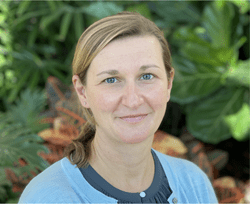
Left photo:
PHOTO DETAILS: (click images for high-res versions)
CREDIT: Amanda Poholek, Ph.D.
CAPTION: Amanda Poholek, Ph.D., assistant professor of pediatrics and immunology at Pitt’s School of Medicine and director of the Health Sciences Sequencing Core at UPMC Children’s Hospital of Pittsburgh.
Right photo:
PHOTO DETAILS: (click images for high-res versions)
CREDIT: Greg Delgoffe, Ph.D.
CAPTION: Greg Delgoffe, Ph.D., associate professor of immunology at Pitt and director of the Tumor Microenvironment Center at UPMC Hillman Cancer Center.


