


9/21/2020
 PITTSBURGH – Researchers at the University of Pittsburgh School of Medicine and Carnegie Mellon University College of Engineering have created a machine-learning algorithm that can detect subtle signs of osteoarthritis—too abstract to register in the eye of a trained radiologist—on an MRI scan taken years before symptoms even begin. These results will publish this week in PNAS.
With this predictive approach, patients could one day be treated with preventative drugs rather than undergoing joint replacement surgery.
 “The gold standard for diagnosing arthritis is x-ray. As the cartilage deteriorates, the space between the bones decreases,” said study co-author Kenneth Urish, M.D., Ph.D., associate professor of orthopaedic surgery at Pitt and associate medical director of the bone and joint center at UPMC Magee-Womens Hospital. “The problem is, when you see arthritis on x-rays, the damage has already been done. It’s much easier to prevent cartilage from falling apart than trying to get it to grow again.”
Right now, the primary treatment for osteoarthritis is joint replacement. And the condition is so prevalent that knee replacement is the most common surgery in the U.S. for people over age 45.
For this study, the researchers looked at knee MRIs from the Osteoarthritis Initiative, which followed thousands of people for seven years to see how osteoarthritis of the knee develops. They focused on a subset of patients who had little evidence of cartilage damage at the beginning of the study.
In retrospect, we now know which of these participants went on to develop arthritis and which didn’t, and the computer can use that information to learn subtle patterns on the MRI scans of presymptomatic people that are predictive of their future osteoarthritis risk.
 “When doctors look at these images of the cartilage, there isn’t a pattern that jumps out to the naked eye, but that doesn’t mean there’s not a pattern there. It just means you can’t see it using conventional tools,” said lead author Shinjini Kundu, M.D., Ph.D., who completed this project as part of her graduate training in the Pitt Medical Scientist Training Program and Carnegie Mellon Department of Biomedical Engineering.
To validate this approach, Kundu, who now is a resident physician and medical researcher at the Johns Hopkins Department of Radiology, trained the model on a subset of the knee MRI data and then tested it on patients it had never seen before. Kundu did this dozens of times, with different participants withheld each time, to test the algorithm on all the data.
Overall, the algorithm predicted osteoarthritis with 78% accuracy from MRIs performed three years before symptom onset.
Currently, there are no drugs that prevent presymptomatic osteoarthritis from developing into full-blown joint deterioration, though there are a few highly effective drugs that can prevent patients from developing a related condition—rheumatoid arthritis.
The goal is to develop the same types of drugs for osteoarthritis. Several candidates already are in the preclinical pipeline.
“Instead of recruiting 10,000 people and following them for 10 years, we can just enroll 50 people who we know are going to be getting osteoarthritis in two or five years,” Urish said. “Then we can give them the experimental drug and see whether it stops the disease from developing.”
Funding for this study was provided by the National Institute of Arthritis and Musculoskeletal and Skin Diseases (grant K08-AR071494).
Additional authors include Beth Ashinsky of Drexel University; Mustapha Bouhrara, Ph.D., and Richard Spencer, M.D., Ph.D., of the National Institute on Aging; Erik Dam, Ph.D., of the University of Copenhagen; Shadpour Demehri, M.D., of Johns Hopkins University; and Mohammad Shifat-E-Rabbi and Gustavo Rohde, Ph.D., of the University of Virginia.
PHOTO INFO: (click images for high-res versions)
PHOTO INFO: (click images for high-res versions)
Top:
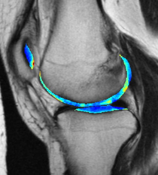
CREDIT: Kundu et al. (2020) PNAS.
CAPTION: Spotting Subtle Patterns In Knee Cartilage: The cartilage in this MRI scan of a knee is colorized to show greater contrast between shades of gray.
Middle:
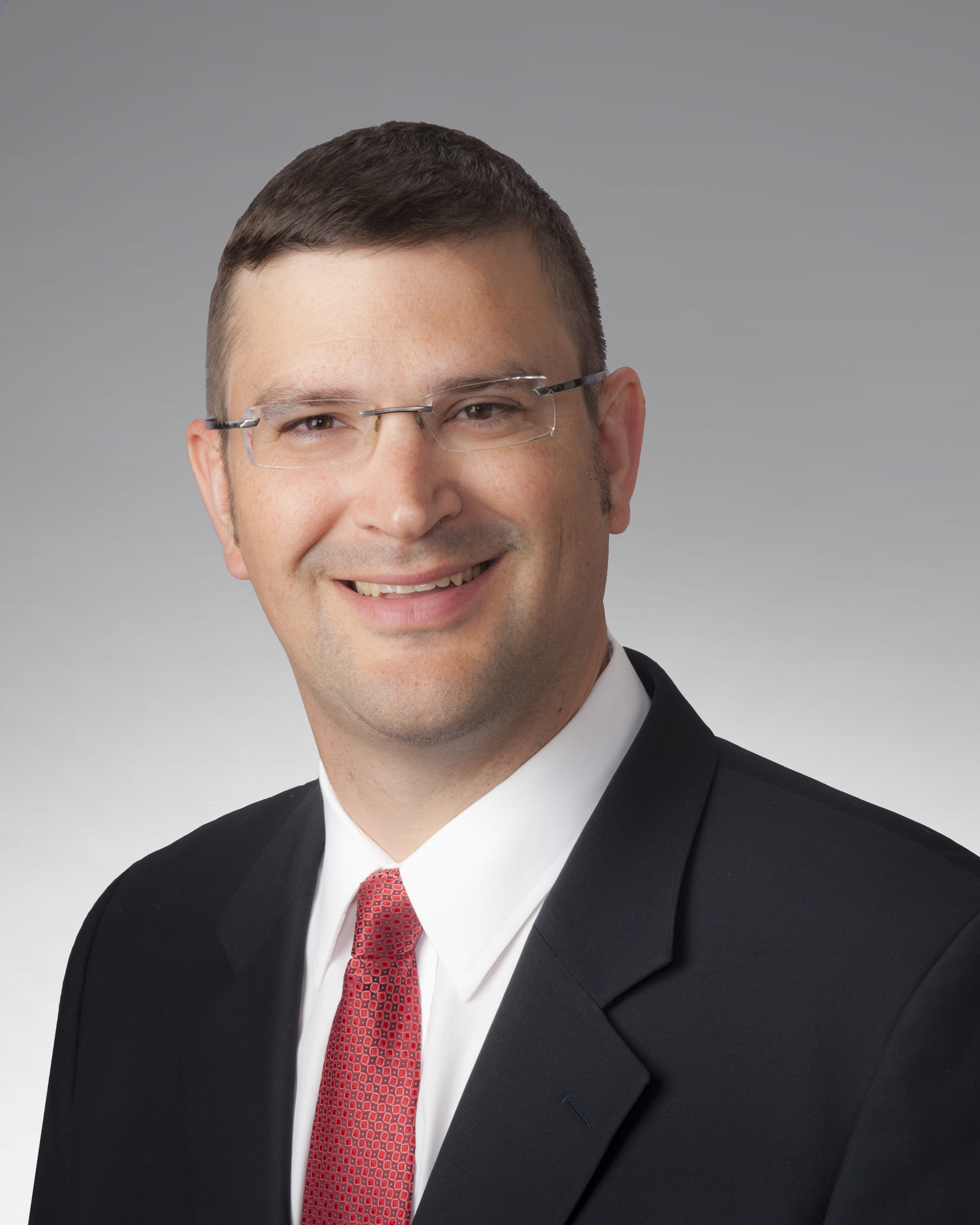
CREDIT: UPMC
CAPTION: Kenneth Urish, M.D., Ph.D., associate professor of orthopaedic surgery at the University of Pittsburgh and associate medical director of the bone and joint center at UPMC Magee-Womens Hospital.
Bottom:
CREDIT: Shinjini Kundu
CAPTION: Shinjini Kundu, M.D., Ph.D., resident physician and medical researcher at the Johns Hopkins Department of Radiology.
