Neuroendoport Clinical Case Study
The Patient
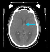
A 41-year-old veteran of Operation Desert Storm had unremitting headaches, confusion, and fainting spells. MRI scans showed a large colloid cyst of the third ventricle.
|
Pre-surgical scan shows a colloid cyst of the third ventricle.
|
|
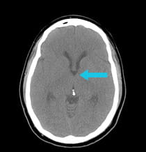
Scan after surgical procedure shows complete removal of colloid cyst.
|
The Challenge & Solution
The colloid cyst was completely removed using the minimally invasive Neuroendoport technique.
The Result
The patient's cognition and headaches improved immediately, and the fainting spells are gone. In the follow-up image, which was performed while the patient was still in the operating room, it is clear that the colloid cyst has been completely removed. Intra-operative imaging with a CT scanner allows UPMC neurosurgeons to confirm safe lesion resection prior to leaving the operating room.
Treatment and results may not be representative of all similar cases.


