Neuroendoport Surgery Clinical Case Study
The Patient
A woman was admitted to the hospital in an acute and severe state of sleepiness with difficulty understanding speech or making words. She also had significant vision loss. MRI scans demonstrated a large mass in the left occipital and temporal lobe region, with a large amount of edema around it.
The Challenge
The glioblastoma was covered with a large amount of functional brain cortex, much of which is critical for speech function. In order to reach the lesion with a standard approach, a large opening in the dura (brain covering) would be required, which would lower her chances of ever regaining speech function.
|
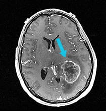
Pre-surgical scan shows 4 cm glioblastoma.
|
|
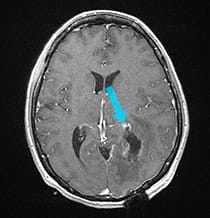
Post-surgical scan shows successful removal following minimally invasive Neuroendoport Surgery.
|
The Solution
Neuroendoport surgery was performed to remove the 4 cm glioblastoma, and the patient improved rapidly, making jokes with the nurses in the recovery room after surgery. Because of the location of the tumor, her visual loss was not salvageable, but her recovery of speech was immediate and profound.
The Result
The patient was able to leave the hospital just two days after surgery. She experienced no significant incisional pain, and was set up for adjuvant radiation and chemotherapy shortly after her discharge.
Treatment and results may not be representative of all similar cases.